
World Pet Obesity · Clinical Assessment Tool
Dog & Cat Muscle Condition Scoring: Standardized Clinical Assessment
Muscle Condition Scoring, or MCS, is a standardized method for evaluating muscle mass in dogs and cats through visual assessment and palpation. Used alongside Body Condition Scoring, or BCS, MCS helps identify muscle loss earlier, supports consistent clinical documentation, and informs treatment and monitoring decisions.
Clinical Context
Why muscle condition is assessed separately from body condition
Body Condition Score estimates body fat. Muscle Condition Score estimates skeletal muscle mass. The two are evaluated independently because they can change in different directions and reflect different underlying processes. A patient may carry excess adipose tissue and still show measurable muscle loss.
Muscle loss may develop with aging, chronic disease, reduced activity, inadequate protein intake, or inflammatory and metabolic conditions. Because loss can progress before it is visually obvious, a structured, repeatable assessment helps teams document change over time and communicate findings consistently.
Key Clinical Principle
Muscle loss can occur at any body condition
Subcutaneous fat can obscure muscle loss on visual inspection alone, so palpation is essential, especially in patients with overweight or obesity. Muscle Condition Score should be recorded alongside, not in place of, Body Condition Score.
Assessment Sites
Where muscle mass is evaluated
Body Condition Score quantifies adiposity but says nothing about lean body mass. In a patient with obesity that distinction matters: excess fat and depleted muscle can coexist in the same animal, and BCS alone will not reveal the muscle loss. Muscle Condition Score was developed to close that gap, evaluating lean mass through visual inspection and palpation of key muscle groups.
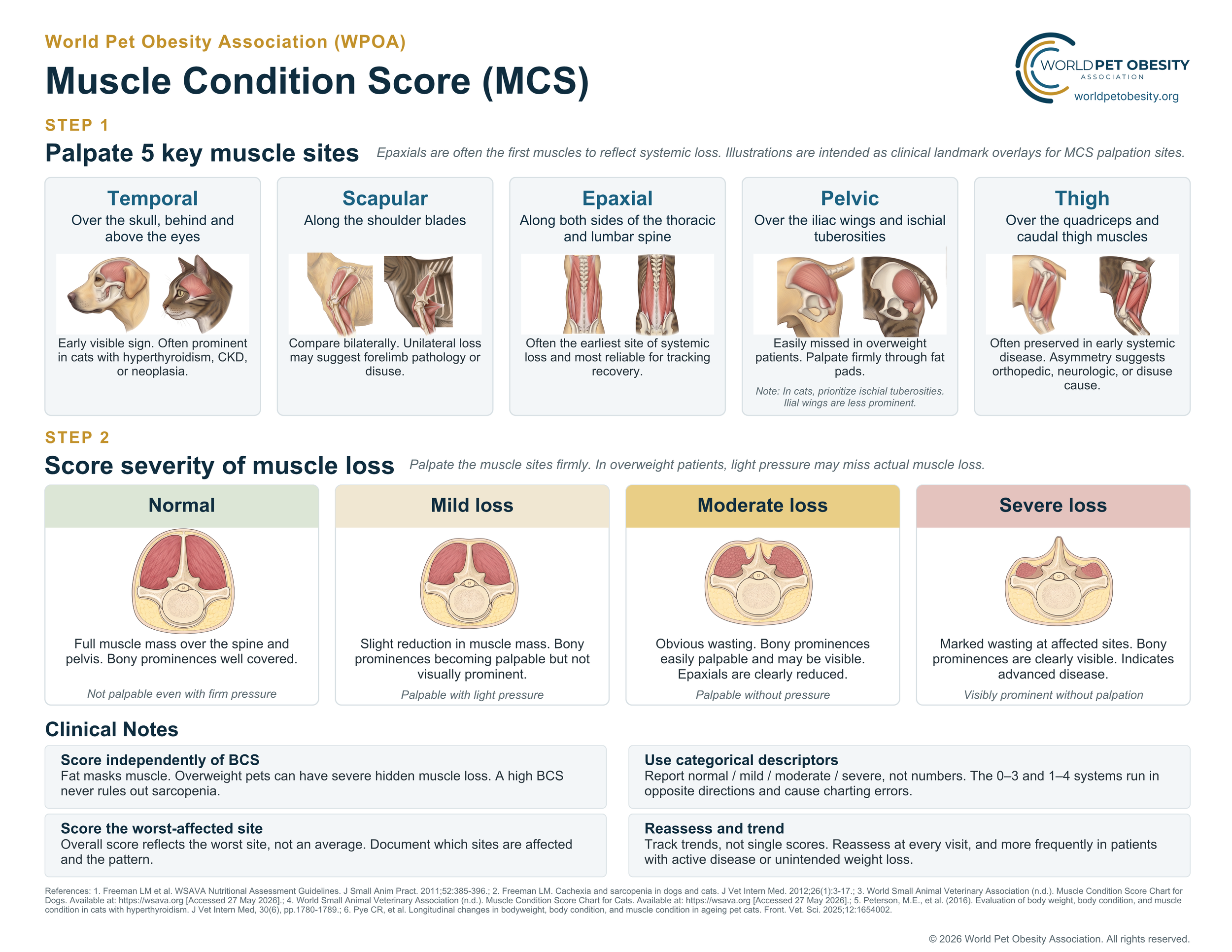
Assessment is performed at five palpation sites where muscle loss is most readily detected: the temporal muscles, the scapular region, the epaxial muscles flanking the thoracic and lumbar vertebrae, the pelvic region (iliac wings and ischial tuberosities), and the thigh (quadriceps and caudal thigh muscles). The overall score reflects the site of greatest muscle loss, not an average across sites.
These five sites matter most in the context of obesity. A patient with excess adiposity can simultaneously lose clinically significant lean mass, a phenotype termed sarcopenic obesity, which BCS and body weight cannot capture. It is especially common in older animals, in whom age-related muscle loss develops in parallel with excess adiposity, and during caloric restriction for weight management, in which preserving lean mass is an explicit therapeutic goal. Because reduced muscle mass is linked to decreased mobility, diminished strength, impaired immune function, and altered metabolism, recognizing it changes how a patient is managed.
Toggle between dog and cat for species-specific illustrations.

Temporal bones
Assess the muscle over the top of the skull. Temporal muscle loss may make the bony contours of the head more prominent.

Scapulae
Palpate over and around the shoulder blades. Reduced muscle coverage makes the scapular spine and surrounding bony contours more apparent.
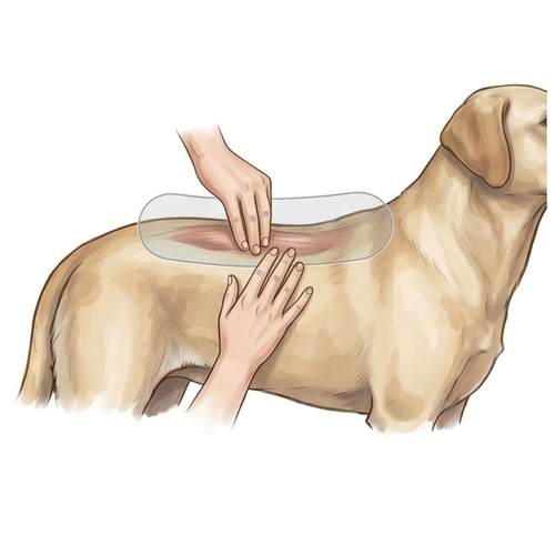
Epaxial muscles
Palpate along the thoracic and lumbar spine. Reduced epaxial muscle makes the dorsal spinous processes and surrounding contours easier to feel.
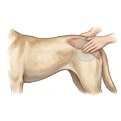
Pelvic bones
Assess muscle coverage over the pelvis and iliac wings. Loss may make pelvic landmarks more visible or more prominent on palpation.
Thigh muscles
Evaluate the hind limb musculature for symmetry, fullness, and loss of contour. Thigh assessment can help identify functional muscle changes.
Score Categories
The four muscle condition grades
Muscle Condition Score is recorded as one of four categories based on findings across assessment sites. Assign a single overall grade from the worst-affected site, not an average across sites, and report it in words rather than numbers. Document which sites are affected and the pattern of loss; sites may not all change at the same rate.
Normal muscle mass
No detectable loss
Palpation: not palpable, even with firm pressure
Full muscle coverage over the temporal region, scapulae, epaxial region, pelvis, and thigh. Bony landmarks are well covered.
Mild muscle loss
Early, subtle change
Palpation: palpable with light pressure
A slight reduction in muscle mass at one or more sites. Bony landmarks are becoming palpable but are not yet visually prominent.
Moderate muscle loss
Readily detectable
Palpation: palpable without pressure
Muscle coverage is clearly reduced, with obvious wasting. Bony landmarks may be visible on inspection, and epaxial muscle is often clearly reduced.
Severe muscle loss
Marked depletion
Palpation: visibly prominent without palpation
Muscle is substantially diminished and bony landmarks are clearly visible. Findings usually indicate advanced disease and warrant further clinical evaluation.
Reference Chart
Printable English MCS reference chart
The complete English Muscle Condition Score chart, including the five palpation sites and the four severity grades, formatted for printing and clinic display. View it below, or download a high-resolution image or PDF.

Performing the Assessment
A consistent, repeatable approach
Muscle Condition Score is most useful when performed the same way at each visit and documented in the medical record alongside Body Condition Score and body weight.
Inspect, then palpate
Begin with visual inspection, then palpate each site. Palpation is more reliable because fat and coat can mask muscle loss.
Work through all sites
Begin at the epaxial muscles, usually the earliest site to reflect systemic loss and the most reliable for tracking change, then assess the temporal region, scapulae, pelvis, and thigh.
Assign an overall grade
Assign a single overall grade from the worst-affected site: normal, mild, moderate, or severe, rather than averaging across sites. Record the category in words, not numbers.
Record alongside BCS
Document both scores together. The pairing gives a fuller picture than either score alone.
Reassess over time
Repeat scoring at routine visits. Trends across visits are often more informative than one measurement.
Investigate change
New or progressing muscle loss may prompt evaluation of diet, activity, pain, aging, and underlying disease.
Interpreting Findings Together
Interpret MCS and BCS together
MCS and BCS describe distinct aspects of the patient. Considered together, they help distinguish excess adiposity from lean tissue status and can improve clinical decision-making.
High BCS with muscle loss
A patient may have obesity and reduced muscle mass. This can change the treatment plan, including protein strategy, activity planning, pain management, and monitoring.
Normal BCS with muscle loss
A patient may appear to have acceptable body condition while losing muscle. This may require investigation for chronic disease, aging changes, reduced activity, or inadequate intake.
Clinical Caution
Do not assume adequate muscle from body weight alone
Stable or increasing body weight does not rule out muscle loss because gains in fat can offset losses in muscle. Palpation across assessment sites remains essential.
Related Resources
Continue with these clinical tools
Body Condition Score Charts
Standardized 9-point body condition tools for dogs and cats supporting adiposity assessment, client communication, and consistency across care and research.
View BCS chartsCalorie & Weight-Loss Calculators
Structured calculation tools to support energy estimates and weight-management planning, interpreted alongside body and muscle condition findings.
Open calculatorsClinical Obesity Framework
A structured approach to defining, diagnosing, and monitoring obesity as a chronic clinical condition in companion animals.
Read the frameworkAbout this tool: The Muscle Condition Score framework is intended for veterinary education and clinical discussion. It should be interpreted alongside Body Condition Score, body weight history, nutrition history, medical history, clinical signs, and a complete physical examination.
About muscle condition scoring
Muscle Condition Score, or MCS, is a standardized assessment used to evaluate skeletal muscle loss in dogs and cats separately from body fat. WPOA provides MCS education, scoring guidance, and downloadable muscle condition score charts in multiple languages to help veterinary teams, educators, researchers, and pet owners recognize normal muscle condition as well as mild, moderate, and severe muscle loss. These multilingual resources support more consistent muscle assessment, clearer documentation, better client communication, and improved monitoring of pets with weight change, aging, chronic disease, overweight, or obesity.